Is It More Than COPD?
Chronic obstructive pulmonary disease (COPD) is a progressive lung disease that causes the airways in the lungs to become inflamed and thicken, which causes the flow of air in and out of the lungs to decrease.1 When this happens, less oxygen gets into your body tissues, making it harder to get rid of the waste gas carbon dioxide. As the disease gets worse, shortness of breath makes it harder to remain active.1,2
According to the Centers for Disease Control and Prevention (CDC), COPD is the third leading cause of death by disease in the United States. More than 16.4 million people have been diagnosed with COPD, but millions more may have the disease without even knowing it. COPD causes serious long-term disability and early death.
There’s no cure for COPD, but treatment can help ease symptoms, lower the chance of complications, and generally improve quality of life. Medications, supplemental oxygen therapy, and surgery are some forms of treatment. Untreated, COPD can lead to a faster progression of disease, heart problems, and worsening respiratory infections.1

Infographic: Boehringer Ingelheim
Signs & Symptoms of COPD
Common symptoms of COPD include breathing difficulty, cough, mucus production and wheezing. People with COPD are also likely to experience episodes called exacerbations, during which their symptoms become worse than usual day-to-day variation and persist for at least several days.1,2
COPD Symptoms include:
- Having to stop and catch your breath during everyday tasks
- A nagging cough, or wheezing sounds
- More mucus than normal, or a change in mucus color or thickness
- Getting tired easily, or feeling sleepy or exhausted
- Frequent respiratory infections (feeling like you always have a cold)

Mucus hypersecretion in all COPD patients affects several important outcomes such as lung function, health-related quality of life, COPD exacerbations, hospitalizations, and mortality.3

The damage caused by COPD comes in multiple forms. The airways leading to the lungs narrow and/or get blocked with mucus. Inside the lungs, air sacs normally take in oxygen and send out carbon dioxide, but in COPD, this tissue breaks down.
With less oxygen for fuel, the systems of the body get weaker, including blood circulation and muscle function. Therefore, patients with COPD must work harder to inhale and exhale and are more prone to feeling tired or short of breath.2
What is Bronchiectasis?
Bronchiectasis is a condition affecting the airways in the lungs that causes cough, increased mucus production, and recurrent lung infections.5 The symptoms are caused by abnormal widening of the airways of the lung, also known as bronchi. The cells lining the airways become inflamed and swollen. These damaged airways can no longer effectively clear mucus and bacteria from the lung. This can lead to flare-ups of cough, mucus production, and shortness of breath.5
Bronchiectasis is caused by one or more infections introduced into the lungs. The most common symptoms are persistent cough and production of excess mucus. Treatments can help reduce coughing, discomfort and mucus production, but this condition is incurable. People with bronchiectasis are more likely to get lung infections. Each lung infection can make the bronchiectasis worse. Therefore, early diagnosis and treatment of bronchiectasis is very important.

Is it COPD or Bronchiectasis?

The prevalence of bronchiectasis is high in patients with moderate-to-severe COPD and it has been associated with exacerbations and bacterial colonization. Some studies have reported a high prevalence of bronchiectasis (42%) in patients with moderate-to-severe COPD.6 Bronchiectasis is hard to differentiate from COPD but recent studies are showing it may be more common than was previously suspected. The wide range in prevalence can be confusing because the symptoms present across many different diagnoses. The clinical overlap between COPD, asthma and bronchiectasis can easily contribute to diagnostic errors.7
Prevalence of bronchiectasis was once thought to be around just 52 cases per 100,000 people, but a 2013 study suggested the actual number is almost triple that amount – it may be closer to 139 per 100,000. In 52 percent of the cases observed, study participants also presented with chronic obstructive pulmonary problems.8
The findings of this study suggest that between 340,000 and 522,000 adults were receiving treatment for bronchiectasis with 70,000 adults newly diagnosed annually, making it more important than ever for medical professionals to recognize the symptoms of Bronchiectasis.8
Both respiratory diseases have symptoms of dyspnea (shortness of breath), chronic cough, potential for daily mucus production, and airflow obstruction. Investigation for bronchiectasis should be considered in patients with COPD with frequent exacerbations (two or more annually) and a previous positive sputum culture for Pseudomonas aeruginosa, which is a bacteria that causes acute and chronic infections.9
COPD is often treated with anti-inflammatory drugs, such as inhaled corticosteroids. However, bronchiectasis often requires an antibacterial approach to stop the vicious cycle of impaired ciliary function leading to bacterial colonization and thick mucus accumulation, leading to inflammation and impairment of airway clearance.9 Diagnosis of bronchiectasis in COPD patients allows for proper treatment of underlying infection and inflammation.
Disorder of the Diaphragm in COPD Patients
A chest X-Ray image of COPD patients may reveal enlarged lungs, a flattened diaphragm, or potentially dangerous air pockets in the lungs. The diaphragm may appear flattened in the chest as a result of hyperinflation as the lungs push against the diaphragm forcing it downward.10 Chronic airflow limitation imposes a load on respiratory muscles as does lung hyperinflation, flattening the diaphragm and reducing its ability to generate tension. Changes in chest wall geometry and diaphragm position are the most recognized and studied mechanisms contributing to respiratory muscle dysfunction.10
COPD patients often develop hyperinflation. Hyperinflation occurs due to expiratory flow limitation caused by reduced lungs’ elastic recoil and increased airway resistance. Hyperinflation increases during exercise and acute exacerbation. Hyperinflation has a significant negative impact on respiratory muscles, particularly the diaphragm.11 Diseases presenting with chronic lung hyperinflation such as COPD are frequently the cause of abnormal diaphragm contractility. Research shows that weak diaphragm muscles can worsen COPD, potentially leading to exacerbations.11

A depressed hemidiaphragm can be the result of asthma, COPD, a tumor or a large pleural effusion. The image depicts flattened diaphragms.
Airway Clearance Therapy

Cough is an effective method of clearing secretions from the larger airways in healthy individuals. However, respiratory muscle impairment leads to difficulty with coughing and clearing secretion, subsequent mucus plugging and pulmonary infection.12 With narrow airways, coughing can have detrimental effects if used inappropriately over an extended period as the primary method of clearing secretions.
Effective mucus clearance is essential for lung health, and airway disease is a consistent consequence of poor clearance.12 Airway clearance techniques (ACTs) to assist with secretion clearance are widely recommended and can include both mucus-mobilizing techniques and assisted cough techniques.13
Studies recommend that ACTs be used as the primary method of mobilizing secretions from the middle and small airways to the larger airways. Then, effective coughing can be used to clear secretions from the larger airways, thereby preserving the integrity of the larger airways.13
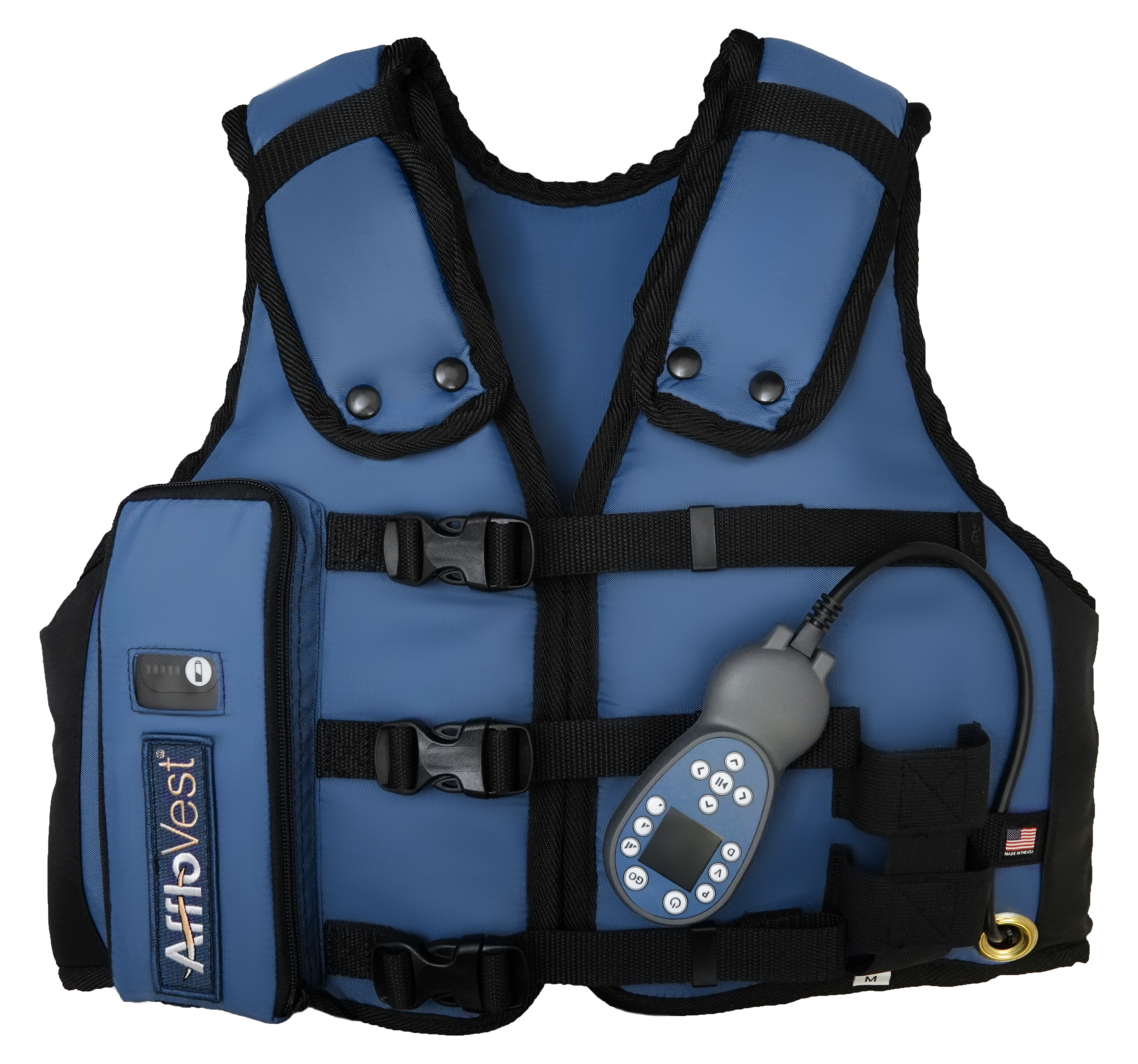
AffloVest Mechanical High Frequency Chest Wall Oscillation HFCWO
The introduction of the AffloVest in 2013 revolutionized the High Frequency Chest Wall Oscillation market by bringing a proven treatment to patients and allowing for mobility during use. The ability for treatment during everyday activities has allowed patients to conduct their therapy while on the go which may led to better, overall patient adherence.
Traditional compression vest systems are comprised of an air-pulse generator and an inflatable vest that is connected by tubes, which leaves patients tethered to a wall during treatment. Once the patient puts the vest on and turns on the generator, each squeeze of the vest provides pressured air pulses to oscillate the chest wall and produce a cough.
AffloVest provides patient-specific therapy with 8 anatomically positioned oscillating motors to create individual pressure waveforms to target all lobes of the lungs, posteriorly and anteriorly, to loosen, thin and mobilize lung secretions. It is designed to mimic the gold standard chest physical therapy (CPT).
The battery powered AffloVest is designed to increase therapy adherence through mobility. The ability to do airway clearance therapy on the go while performing normal, daily activities can provide more consistent therapy compliance and an improved quality of life.
REFERENCES
1. Lung https://www.lung.org/lung-health-diseases/lung-disease-lookup/copd/learn-about-copd
2. COPD Foundation: https://www.copdfoundation.org/What-is-COPD/Understanding-COPD/What-is-COPD.aspx
3. Ramos, F. et al. Clinical Issues of Mucus Accumulation in COPD. International Journal of COPD. 2014.
4. Cook, N.S. et al. Patients’ Perspectives on COPD: Findings from a Social Media Listening Study. European Respiratory Journal Open Research. 2019
5. American Lung Association: https://www.lung.org/lung-health-and-diseases/lung-disease-lookup/bronchiectasis/
6. Kosmas E, et al., Bronchiectasis in Patients with COPD: An Irrelevant Imaging Finding or a Clinically Important Phenotype? CHEST 2016;150(4):894A.
7. Aksamit, T. et al. Bronchiectasis and Chronic Airway Disease: It is not just about Asthma and COPD. CHEST Journal. October 2018
8. Weycker D, et al., Prevalence and Incidence of Non-cystic Fibrosis BE Among US Adults in 2013. Chron Respir Dis 2017. Nov;14(4): 377-384.
9. Athanazio, R et al. Airway disease: similarities and differences between asthma, COPD and bronchiectasis. Clinics (2012) 67(11):1335
10. Alana Biggers, MD, MPH Does COPD show up on an X-ray? https://www.medicalnewstoday.com/articles/323363
11. Sarkar M, Bhardwaz R, Madabhavi I, Modi M. Physical signs in patients with chronic obstructive pulmonary disease. Lung India. 2019;36(1):38–47.
12. Fahy JV, Dickey BF. Airway mucus function and dysfunction. N Engl J Med. 2010;363(23):2233–2247.
13. McIlwaine M, Bradley J, Elborn JS, et al. Personalizing airway clearance in chronic lung disease. Eur Respir Rev 2017; 26: 16008